Doctors print sick organs and only plan operations. New technologies offer extraordinary opportunities
Holomedicine, augmented reality and 3D printing – thanks to these technologies, doctors can plan operations precisely as never before. It is also a new path for students who can gain their first touches on models and phantoms, not on patients.
Dr. hab. n. med. Paweł Rynio, specialist in vascular surgery, is the originator and manager of the first 3D Medical Technology Center in Poland at the Independent Public Clinical Hospital No. 2 in Szczecin. The resort has been operating since October 2022. It was created under the Interreg VA Cooperation Program Mecklenburg-Vorpommern/Brandenburg/Poland under the “European Territorial Cooperation” objective of the European Regional Development Fund (ERDF). The project partner is the Medical University of Greifswald.
Katarzyna Świerczyńska, “Wprost”: A patient comes in, you diagnose him with an aortic aneurysm, and then you print the aorta with the aneurysm?
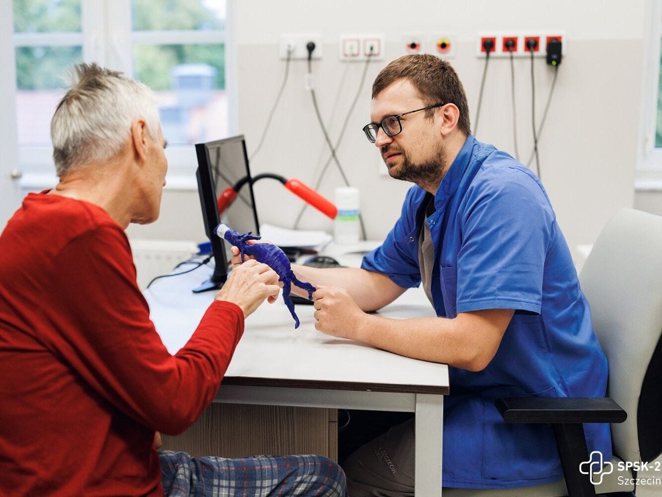
Pawel Rynio: Yes. We do it to show the patient what this pathology looks like, in this case an aneurysm. Thanks to this, we can explain very understandably to the patient what awaits him, what the procedure will look like.
But we also make such a 3D print for ourselves, for doctors, when the operation to remove an aortic aneurysm needs to be personalized. 3D printing is particularly useful when the aneurysm has an unusual structure and a special, tailor-made prosthesis is required, i.e. the so-called stent-graft.
We use it to replace the altered section of the aorta, in other words, an artificial vascular prosthesis. Printing a template, i.e. the patient’s anatomy, and then applying a standard prosthesis to it allows for its personalization and adaptation to a specific patient. The arrangement of the vessels is very individual, they run a little differently in everyone, differ in size, which is why it is so important.
What do patients say when they see their aneurysm and can touch it?
They say: wow! They are literally in shock. And it always dispels the doubts of those who are not convinced whether it makes sense to operate. When a man sees with his own eyes how big it is, can touch it, he is fully aware of the problem we are dealing with. He sees what the consequences can be if an aneurysm ruptures. Importantly, we are also able to use this method in emergency cases.
What the possibility of 3D printing changes from the doctor’s perspective. You mentioned the possibility of personalizing the prosthesis. But does the fact that the doctor picks up a printout identical to what the patient has inside help in the operation itself?
Of course. We use 3D printing quite often to consider the treatment strategy and intraoperative management.
When you opened the center, prof. Maciej Patryk z of the Medical University of Greifswald gave specific data. He said that thanks to 3D printing, even 13-20 percent. cases where, with traditional imaging methods, doctors considered a given case inoperable, changed their minds after seeing the model. Is this actually happening?
There are some things we cannot see or imagine on the basis of two-dimensional scans. In such difficult cases, we take a model and discuss, looking for a solution.
This is very often useful in urology, because of course we cooperate with various departments of our hospital. Kidney tumors are very problematic and thanks to the printout we are able to plan a precise and possibly the most kidney-sparing operation. We are talking here about cases when the preservation of part of the kidney is considered, i.e. partial nephrectomy.
The doctor sees exactly how all the vessels that approach the tumor run. We recently prepared a very peculiar anatomy. The patient had cancer, but at the same time he had kidneys connected to each other. He was born with such a defect that instead of two kidneys on either side of his loins, he had one, fused. Such a case is a conglomerate of everything – unusual arrangement of arteries, veins, ureters. And then there was cancer. 3D printing turned out to be invaluable in planning this unusual operation.
Have you changed your plans after seeing the printout?
Yes, we are talking about changing the operating strategy here. For example, we initially qualify the patient for endovascular surgery, and then, after seeing the model, it turns out that this type of surgery will absolutely fail and we decide, for example, on open surgery. This is often the case with these complex pathologies.
Do you print before each operation?
Printing makes no sense in standard, obvious cases. This method, as I said, is especially useful in cases of atypical anatomy or procedures that are rarely performed.
You are the first hospital in Poland that has such a possibility.
We cooperate with all surgical departments in our hospital and I regret that we do not have orthopedics or, for example, cranial surgery, because these are the fields of medicine where working with 3D models is particularly useful and this technology is already successfully used around the world.
Every hospital should have a center like this?
At least there could be larger consortia where several hospitals could take advantage of this possibility. In the United States, 20 percent of hospitals already have hospital 3D printing laboratories, so we still have a lot to do in this matter.
Your center is not only 3D prints, but also holomedicine. It sounds like something out of a science fiction movie, but I myself had the opportunity to put on special goggles and see models of specific organs in front of me. It’s impressive, but how does it work in practice?
Holomedicine allows you to visualize specific anatomy details in 3D without the need for 3D printing. It has its pluses and minuses. The pluses are that we don’t use any materials, we don’t waste time on printing, but the minuses – unfortunately, the level of these holograms is not as good as in the case of printouts, there is still a lot to do here.
I would say holomedicine is in its infancy. But I’m sure in the future the use of good quality holograms and mixed reality will be something we’ll be using a lot.
Because imagine that the doctor puts on the goggles and can see the patient’s anatomy in all its details, every pathology. I think it takes at least a few more years to make it possible.
Students also use the center. The gentleman, learning his profession, did not have such opportunities.
This is a great thing for students, because they can see pathologies in the form of a 3D print or hologram. They can touch, see, turn from any side. Anyway, this direction related to education is very important to us. We want to obtain further funding from funds for cross-border projects and create a Laboratory of Surgical Skills, the so-called “Skill-Lab” equipped with special trainers for students, where they will be able to practice operations, e.g. laparoscopic or endoscopic, not on patients, but on models, on phantoms. Such a training center will combine holomedicine with virtual reality trainers. We want to do this together with our partners from Greisfwald.
What is the future related to the use of 3D in medicine, holomedicine and augmented reality?
This will primarily lead to the personalization of treatment, but also to better education of students who will take their first steps on phantoms, not on people. I hope that soon we will perform operations in mixed reality goggles, which will precisely display to us in the operating field where specific anatomical structures are located. I think this is the future.